Digital Microscope Imager

pH meter

Camera, Telescope & Optic

Kindle Wireless Reading Device. Slim and lightweight - only 8.5 ounces

Pocket-Sized Sensor for Detect 'Date Rape' Drugs, A New Line of Defense Against Sexual Assault

Saturday, August 20, 2011


Date rape can be experienced by anyone, at any time. It's always better to be cautious than sorry. Not all sexual attackers are strangers who jump you in the streets. Sexual assaults can take place during social interactions such as dates while you are out with someone you know. 

In most cases of 'date rape', alcohol is the substance used to assist in forcing non-consensual sex from a partner. In other cases, drinks may also be 'spiked' by date rape drugs such as GHB, rohypnol and ketamine so that resistance is neutralised or to render the victim unconscious.

Smart women know it's wise to beware when out at a bar or club -- there could be more than just alcohol in that cocktail. Psychoactive substances classified as "date rape" drugs can be dropped into an unsuspecting victim's drink, rendering her barely conscious and susceptible to sexual assault.


What are date rape drugs?
These are drugs that are sometimes used to assist a sexual assault. Sexual assault is any type of sexual activity that a person does not agree to. It can include touching that is not okay; putting something into the vagina; sexual intercourse; rape; and attempted rape. These drugs are powerful and dangerous. They can be slipped into your drink when you are not looking. The drugs often have no color, smell, or taste, so you can't tell if you are being drugged. The drugs can make you become weak and confused -- or even pass out -- so that you are unable to refuse sex or defend yourself. If you are drugged, you might not remember what happened while you were drugged. Date rape drugs are used on both females and males.


The three most common date rape drugs are:

Rohypnol (roh-HIP-nol). Rohypnol is the trade name for flunitrazepam (FLOO-neye-TRAZ-uh-pam). Abuse of two similar drugs appears to have replaced Rohypnol abuse in some parts of the United States. These are: clonazepam (marketed as Klonopin in the U.S. and Rivotril in Mexico) and alprazolam (marketed as Xanax). 

Rohypnol is also known as: Circles, Forget Pill, LA Rochas, Lunch Money, Mexican Valium, Mind Erasers, Poor Man's Quaalude, R-2, Rib, Roach, Roach-2, Roches, Roofies, Roopies, Rope, Rophies, Ruffies, Trip-and-Fall, Whiteys.

GHB, which is short for gamma hydroxybutyric (GAM-muh heye-DROX-ee-BYOO-tur-ihk) acid. GHB is also known as: Bedtime Scoop, Cherry Meth, Easy Lay, Energy Drink, G, Gamma 10, Georgia Home Boy, G-Juice,ok, Goop, Great Hormones, Grievous Bodily Harm (GBH), Liquid E, Liquid Ecstasy, Liquid X, PM, Salt Water, Soap, Somatomax, Vita-G.

Ketamine (KEET-uh-meen), also known as: Black Hole, Bump, Cat Valium, Green, Jet, K, K-Hole, Kit Kat, Psychedelic Heroin, Purple, Special K, Super Acid.

Possessing both sedative and amnesiac effects, date rape drugs are increasingly slipped into drinks at parties, clubs and bars. With rates of drug-assisted sexual assault growing around the world, it's a dangerous social problem in desperate need of a solution. According to the U.S. Department of Justice, some 200,000 women were raped in the US in 2007 with the aid of a date rape drug -- and because so many cases go unreported, the actual number is believed to be 80 to 100 percent higher.

Until now, the researchers explain, real time date rape drug detection has been impossible. No sensor sensitive enough to detect the drugs had been developed, and after a few hours, the drugs become undetectable in the human bloodstream, making their presence difficult to prove.

Drug detection in one sip

Prof. Fernando Patolsky and Dr. Michael Ioffe of Tel Aviv University's Sackler Faculty of Exact Sciences have developed an easy-to-use sensor that, when dipped into a cocktail, will instantly detect the presence of a date rape drug. When ready for commercial purchase in just a few years, the sensor will be lightweight and discreet, easily transportable in a pocket or purse.

The researchers say that the sensor can detect GHB and ketamine, the most commonly used date rape drugs, with 100 percent accuracy. The technology was recently presented at the Nano Conference 2011 in Israel.

The new system works on simple optics principles, says Prof. Patolsky. Though date rape drugs are effective because they're colorless and tasteless when mixed into a cocktail, they do subtly change the optical properties of the drink. When a ray of light comes into contact with a drugged drink, a "signal change" occurs and the sensor sounds the alarm, which could be a beeping noise or a small flashing light in environments that are dark and loud.

To test the accuracy of the sensor, Prof. Patolsky and Dr. Ioffe had bartenders prepare a large number of the 15 most popular cocktails. Fifty of these drinks were randomly spiked with GHB, without the researchers' knowledge. When their test was conducted, each of the spiked drinks was correctly identified, and there were no false positives.

Only a tiny "sip" of one to ten microliters is required for the sensor to detect the presence of a date rape drug, Prof. Patolsky says.


Affordable personal protection

Researchers are now working on miniaturizing the system, making it easy and affordable for personal use. Each device, says Prof. Patolsky, might look like a pen or clip, easy to dip into a glass. A disposable cartridge inside, responsible for recognizing the presence of a drug, would be able to identify two to three spiked drinks before needing to be replaced -- and new cartridges would each cost under a dollar.

Dr. Ioffe is also hoping to widen the range of drugs that the sensor can correctly identify. "Currently," he says, "the system is geared towards detecting GHB and ketamine. We hope to expand the system so it will identify additional date rape drugs as well."

Reference Book: Nanotechnology & Biosensor

Nanotechnology Enabled In situ Sensors for Monitoring HealthDrug-Facilitated Sexual Assault: A Forensic Handbook3D Cell-Based Biosensors in Drug Discovery Programs: Microtissue Engineering for High Throughput Screening
Nanotechnology Enabled In situ Sensors for ...
$102.77
Drug-Facilitated Sexual Assault: A Forensic...    
$107.00
3D Cell-Based Biosensors in Drug Discovery ...
by William S. Kisaalita
$111.65

NanoScience in BiomedicineNanomedicine Design of Particles, Sensors, Motors, Implants, Robots, and Devices (Engineering in Medicine & Biology)Fluorescence Sensors and Biosensors
NanoScience in Biomedicine $172.01Medicine Design of Particles, Sensors, ...
$142.34
Fluorescence Sensors and Biosensors
$134.13


Defense Against Bioterror: Detection Technologies, Implementation Strategies and Commercial Opportunities--Proceedings of the NATO Advanced Research Workshop on Defense against Bioterror held in Madrid, Spain from 8 to 11 April 2004 (NATO Security through Science, Series B: Physics and Biophysics, Vol. 1)Biomedical NanotechnologyTiO2 Nanotube Arrays: Synthesis, Properties, and Applications
Defense Against Bioterror: Detection Techno... 
$169.00
Biomedical Nanotechnology
$157.95                                  
TiO2 Nanotube Arrays: Synthesis, Properties...
by Craig A. Grimes
$194.00

Nanotechnology

Monday, May 2, 2011


Nanotechnology is science at the size of individual atoms and molecules: objects and devices measuring mere billionths of a meter, smaller than a red blood cell. Nanotechnology is rapidly becoming an interdisciplinary field. Biologists, chemists, physicists and engineers are all involved in the study of substances at the nanoscale.

Nanoscale
Experts sometimes disagree about what constitutes the nanoscale, but in general, you can think of nanotechnology dealing with anything measuring between 1 and 100 nm. Larger than that is the microscale, and smaller than that is the atomic scale.

As small as a nanometer is, it's still large compared to the atomic scale. An atom has a diameter of about 0.1 nm. An atom's nucleus is much smaller -- about 0.00001 nm. Atoms are the building blocks for all matter in our universe. You and everything around you are made of atoms. Nature has perfected the science of manufacturing matter molecularly. For instance, our bodies are assembled in a specific manner from millions of living cells. Cells are nature's nanomachines.

At the atomic scale, elements are at their most basic level. On the nanoscale, we can potentially put these atoms together to make almost anything. In a lecture called "Small Wonders: The World of Nanoscience," Nobel Prize winner Dr. Horst Störmer said that the nanoscale is more interesting than the atomic scale because the nanoscale is the first point where we can assemble something -- it's not until we start putting atoms together that we can make anything useful.

One of the exciting and challenging aspects of the nanoscale is the role that quantum mechanics plays in it. At nanoscale, materials have different chemical and physical properties than those of the same materials in bulk, because quantum mechanics is more important. The rules of quantum mechanics are very different from classical physics, ­which means that the behavior of substances at the nanoscale can sometimes contradict common sense by behaving erratically.
For example, carbon atoms can conduct electricity and are stronger than steel when woven into hollow microscopic threads. You can't walk up to a wall and immediately teleport to the other side of it, but at the nanoscale an electron can -- it's called electron tunneling. Substances that are insulators in bulk form, meaning that they can't carry an electric charge,  might become semiconductors when reduced to the nanoscale. Melting points can change due to an increase in surface area. Much of nanoscience requires that you forget what you know and start learning all over again.

So what does this all mean? Right now, it means that scientists are experimenting with substances at the nanoscale to learn about their properties and how we might be able to take advantage of them in various applications. Engineers are trying to use nano-size wires to create smaller, more powerful microprocessors. Doctors are searching for ways to use nanoparticles in medical applications. Still, we've got a long way to go before nanotechnology dominates the technology and medical markets.

At this time, nanoparticles are already widely used in certain commercial consumer products, such as suntan lotions, "age-defying" make-up, and self-cleaning windows that shed dirt when it rains. One company manufactures a nanocrystal wound dressing with built-in antibiotic and anti-inflammatory properties.

On the horizon is toothpaste that coats, protects and repairs damaged enamel, as well as self-cleaning shoes that never need polishing. Nanoparticles are also used as additives in building materials to strengthen the walls of any given structure, and to create tough, durable, yet lightweight fabrics.


Reference Books: Nanotechnology

Nano-OptoelectronicsFoundations of NanomechanicsNanostructuring Operations in Nanoscale Science and EngineeringNanocomputing: Computational Physics for Nanoscience and NanotechnologyNano-Optics
Nano-Optoelectronics

Foundations of Nanomechanics
by Andrew N. Cleland
Nanostructuring Operations in Nanoscale Science and Engineering
by Kal Sharma
Nano-Optics
by James Hsu
Foundations of Nanomechanics


Biosensors

Sunday, January 23, 2011


What Is a Biosensor?

A biosensor consists of two components: a bioreceptor and a transducer. The bioreceptor is a biomolecule that recognizes the target analyte, and the transducer converts the recognition event into a measurable signal. The uniqueness of a biosensor is that the two components are integrated into one single sensor (Fig1). 

Fig 1. Biosensor Configuration. Biosensor = bioreceptor + transducer
This combination enables one to measure the target analyte without using reagents. For example, the glucose concentration in a blood sample can be measured directly by a biosensor made specifically for glucose measurement, by simply dipping the sensor in the sample. This is in contrast to the commonly performed assays, in which  many sample preparation steps are necessary and each step may require a reagent to treat the sample. The simplicity and the speed of measurements that require no specialized laboratory skills are the main advantages of a biosensor.

Enzyme is a Bioreceptor.

When we eat food such as hamburgers and french fries, it is broken down into small molecules in our body via many reaction steps (these breakdown reactions are called catabolism). These small molecules are then used to make the building blocks of our body, such as proteins (these synthesis reactions are called anabolism). Each of these catabolism and anabolism reactions (the combination is called metabolism) are catalyzed by a specific enzyme. Therefore, an enzyme is capable of recognizing a specific target molecule (Figure 2). This biorecognition capability of the enzyme is used in biosensors. Other biorecognizing molecules (= bioreceptors) include antibodies, nucleic acids, and receptors.
Fig 2. Specivity of biosensor. TR=Transducer

Immobilization of Bioreceptor

One major requirement for a biosensor is that the bioreceptor be immobilized in the vicinity of the transducer. The immobilization is done either by physical entrapment or chemical attachment. Chemical attachment often involves covalent bonding to transducer surface by suitable reagents. It is to be noted that only minute quantities of bioreceptor molecules are needed, and they are used repeatedly for measurements.

Transducer


A transducer should be capable of converting the biorecognition event into a measurable signal (Figure 3). Typically, this is done by measuring the change that occurs in the bioreceptor reaction. For example, the enzyme glucose oxidase is used as a bioreceptor in a glucose biosensor that catalyzes the following reaction:

Fig 3. Three possible transducers for glucose measurement.


 To measure the glucose in aqueous solutions, three different transducers can be used:
  1. An oxygen sensor that measures oxygen concentration, a result of glucose reaction 
  2. A pH sensor that measures the acid (gluconic acid), a reaction product of glucose 
  3. A peroxidase sensor that measures H2O2 concentration, a result of glucose reaction
Note that an oxygen sensor is atransducer that converts oxygen concentration into electrical current. A pH sensor is a transducer that converts pH change into voltage change. Similarly, a peroxidase sensor is a transducer that converts peroxidase concentration into an electrical current.

Biosensor Characteristics


Biosensors are characterized by eight parameters. These are: 
  1. Sensitivity is the response of the sensor to per unit change in analyte concentration.
  2. Selectivity is the ability of the sensor to respond only to the target analyte. That is, lack of response to other interfering chemicals is the desired feature.
  3. Range is the concentration range over which the sensitivity of the sensor is good. Sometimes this is called dynamic range or linearity.
  4. Response time is the time required for the sensor to indicate 63% of its final response due to a step change in analyte concentration.
  5. Reproducibility is the accuracy with which the sensor’s output can be obtained.
  6. Detection limit is the lowest concentration of the analyte to which there is a measurable response.
  7. Life time is the time period over which the sensor can be used without significant deterioration in performance characteristics.
  8. Stability characterizes the change in its baseline or sensitivity over a fixed period of time. 

Considerations in Biosensor Development


Once a target analyte has been identified, the major tasks in developing a biosensor involve:
  1. Selection of a suitable bioreceptor or a recognition molecule.
  2. Selection of a suitable immobilization method.
  3. Selection and design of a transducer that translates binding reaction into measurable signal.
  4. Design of biosensor considering measurement range, linearity, and minimization of interference, and enhancement of sensitivity.
  5. Packaging of the biosensor into a complete device.
The first item above requires knowledge in biochemistry and biology, the second and third require knowledge in chemistry, electrochemistry and physics, and the fourth requires knowledge of  kinetics and mass transfer. Once a biosensor has been designed, it must be packaged for convenient manufacturing and use. The current trend is miniaturization and mass production. 

Modern IC (integrated circuit) fabrication technology and micromachining technology are used increasingly in fabricating biosensors, as they reduce manufacturing costs. Therefore, an interdisciplinary research team, consisting of the various disciplines identified above, is essential for successful development of a biosensor.

Biosensor References:

Biosensors: Theory and Applications
Biosensors: Theory and Applic...
Principles of Bacterial Detection: Biosensors, Recognition Receptors and Microsystems
Principles of Bacterial Detection
Mathematical Modeling of Biosensors: An Introduction for Chemists and Mathematicians (Springer Series on Chemical Sensors and Biosensors)
Mathematical Modeling of Biosensors

Engineering Biosensors: Kinetics and Design Applications
Engineering Biosensors: Kinetics..
Biosensors (The Practical Approach Series)
Biosensors (The Practical.....)
Electrochemical Sensors, Biosensors and their Biomedical Applications

Application of Ultrasound

Thursday, December 16, 2010


Ultrasound has many applications in our lifes. Below are some of the applications.

Medical
Ultrasound has a broad range of applications in medicine, where it is referred to as medical ultrasound.
It is widely used in obstetrics to follow the development of the fetus during pregnancy, in cardiology
where images can display the dynamics of blood flow and the motion of tissue structures (referred to as
real-time imaging), and for locating tumors and cysts. 3-D imaging, surgical applications, imaging from
within arteries (intravascular ultrasound), and contrast imaging are among the newer developments.


Ultrasonic leak detector

Industrial
In industry, ultrasound is utilized for examining critical structures, such as pipes and aircraft fuselages, for cracks and fatigue. Manufactured parts can likewise be examined for voids, flaws, inclusions, debonding, etc. Such defects can exist immediately after manufacturing, or were formed due to stresses, corrosion, etc. Ultrasound has also widespread use in process control. The applications are collectively called Non- Destructive Testing (NDT) or Non-Destructive Evaluation (NDE). In addition, acoustic microscopy refers
to microscopic examinations of internal structures that cannot be studied with a light microscope, such as an integrated circuit or biological tissue.

Underwater
Ultrasound is likewise an important tool for locating structures in the ocean, such as wrecks, mines, submarines, or schools of fish; the term SONAR (SOund Navigation And Ranging) is applied to these applications.

Range Measurements, Air
Ultrasound range measurements are used in cameras, in robotics, for determining dimensions of rooms, etc. Measurement frequencies are typically around 50 kHz to 60 kHz. The measurement concept is pulse-echo, but with burst excitation rather than pulse excitation. Special electronic circuitry and a thin low-acoustic-impedance air transducer is most commonly used. Rugged solid or composite piezoelectric-based transducers, however, can also be used, sometimes up to about 500 kHz.
  
Thickness Measurement for Testing, Process Control, Etc.
Measurement of thickness is a widely used application of ultrasound. The measurements can be done with direct coupling between the transducer and the object of interest, or — if good surface contact is difficult to establish — with a liquid or another coupling agent between the transducer and the object. Ultrasound measurements of thickness have applications in process control, quality control, measuring build-up of ice on an aircraft wing, detecting wall thickness in pipes, as well as medical applications. The instrumentation involves a broadband transducer, pulser-receiver, and display or, alternatively, echo detecting circuitry and numerical display.

Ultrasonic Flow Sensor
Doppler Flow Measurements
The flow velocity of a liquid or a moving surface can be determined through Doppler measurements, provided that the liquid or the surface scatters ultrasound back in the direction of the transducer, and that the angle between the flow direction and the ultrasound beam is known. Further details are given in the section about Doppler processing. CW and PW Doppler instruments are commercially available, with CW instrumentation being by far the least expensive.

Upstream/Downstream Volume Flow Measurements
When flow velocity is measured in a pipe with access to one or both sides, an ultrasound transmission technique can be used in which transducers are placed on the same or opposite sides of the pipe, with one transducer placed further upstream than the other transducer. From the measured difference in travel time between the upstream direction and the downstream direction, and knowledge about the pipe geometry, the volume flow can be determined. Special clamp-on transducers and instrumentation are available. An overview of flow applications in NDE is given in.

Elastic Properties of Solids
Since bulk sound speed varies with the elastic stiffness of the object, sound speed measurements can be used to estimate elastic properties of solids under different load conditions and during solidification processes. Such measurements can also be used for measurement of product uniformity and for quality assurance. The measurements can be performed on bulk specimens or on thin rods, using either pulse-echo or transmission instrumentation. Alternatively, measurements of the material’s own resonance frequencies can be performed for which commercial instruments, such as the Grindo-sonics, are available.

Porosity, Grain Size Estimation

Measurement of ultrasound attenuation can reveal several materials parameters. By observing the attenuation in metals as a function of frequency, the grain size and grain size distribution can be estimated. Attenuation has been used for estimating porosity in composites. In medical ultrasound, attenuation is widely used for tissue characterization, that is, for differentiating between normal and pathological tissues. Pulse-echo instrumentation interfaced with a digitizer and a computer for data analysis is required.

Acoustic Microscopy
The measurement approaches utilized in acoustic microscopy are similar to other ultrasound techniques, in that A-scan, B-scan, and C-scan formats are used. It is in the applications and the frequency ranges where acoustic microscopy differs from conventional pulse-echo techniques. Although acoustic microscopes have been made with transducer frequencies up to 1 GHz, the typical frequency range is 20 MHz to 100 MHz, giving spatial resolutions in the range from 100 µm to 25 µm. Acoustic microscopy is used for component failure analysis, electronic component packaging, and internal delaminations and disbonds in materials, and several types of acoustic microscopes are commercially available.


Ultrasonic Reference Books:

Ultrasonics: Data, Equations and Their Practical Uses
Ultrasonics: Data, Equations and Their Prac...
Physical Principles of Medical Ultrasonics
Fundamentals and Applications of Ultrasonic Waves (Pure and Applied Physics)
Fundamentals and Applications of Ultrasonic...

ULTRASONIC SENSOR

Friday, November 26, 2010


Ultrasonic sensing techniques have become mature and are widely used in the various fields of engineering and basic science. Actually, many types of conventional ultrasonic instruments, devices and sophisticated software are commercialized and used for both industrial and medical applications. One of advantages of ultrasonic sensing is its outstanding capability to probe inside objectives nondestructively because ultrasound can propagate through any kinds of media including solids, liquids and gases except vacua.

In typical ultrasonic sensing the ultrasonic waves are travelling in a medium and often focused on evaluating objects so that a useful information on the interaction of ultrasonic energy with the objects are acquired as ultrasonic signals that are the wave forms variations with transit time. Such ultrasonic data provides the fundamental basis for describing the outputs of ultrasonic sensing and evaluating systems.

Features of Ultrasonic Waves

Ultrasound is an acoustic wave with a frequency higher than the audible range of the human ear, which is 20 kHz. Ultrasound can be within the audible range for some animals, like dogs, bats, or dolphins. In the years around 1883, Sir Francis Galton performed the first known experiments with whistles generating ultrasound. Many decades later, people started to find ultrasound applications in engineering, medicine, and daily life.

The basic principle for the use of ultrasound as a measurement tool is the time-of-flight technique. The pulse-echo method is one example. In the pulse-echo method, a pulse of ultrasound is transmitted in a medium. When the pulse reaches an another medium, it is totally or partially reflected, and the elapsed time from emission to detection of the reflected pulse is measured. This time depends on the distance and the velocity of the sound. When sound travels with a known velocity c, the time t elapsed between the outgoing signal and its incoming echo is a measure of the distance d to the object causing the echo.

It is known that frequency range of sound audible to humans is approximately 20–20,000Hz (cycles per second). Ultrasound is simply sound that are above the frequency range of human hearing. When a disturbance occurs at a portion in an elastic medium, it propagates through the medium in a finite time as a mechanical sound wave by the vibrations of molecules, atoms or any particles present. Such mechanical waves are also called elastic waves. Ultrasound waves or ultrasonic waves are the terms used to describe elastic waves with frequency greater than 20,000Hz and normally exist in solids, liquids, and gases.

It is important to understand the behaviour and properties of ultrasonic waves in media, to design ultrasonic sensors and develop ultrasonic sensing systems. Some basic features of ultrasonic waves are introduced here.

Types of Wave (Modes of Propagation)

What types of ultrasonic waves can exist? The answer to this question can basically be given from solutions of the wave equations that predict wave behaviours by showing that material properties and body shape dictate the vibrational response to the applied forces that drive the wave motion.  In short, there are two types of ultrasonic waves: bulk (fundamental) waves that propagate inside of anobject, and guided waves that propagate near the surface or along the interface of an object.

Waves that propagate wholly inside an object, independent of its boundary and shape, are called bulk waves. Two types of bulk waves can exist in an isotropic medium: longitudinal (or dilatational, compression, primary), and shear (or distortional, transverse, secondary) waves as shown schematically in Fig. 1.

Ultrasonic wave propagations are usually described in terms of the direction of particles motion in relation to the direction in which the wave propagates. The longitudinal waves can be defined on this basis as waves in which the particle motion is parallel to the direction of the wave propagation. 

The shear waves are defined as waves in which the particle motion is perpendicular to the direction of the propagation. Both waves can exist in solids because solids, unlike liquids and gasses, have rigidity that is a resistance to shear aswell as compressive loads. However, the shear waves cannot exist in liquids and gasses because of no resistance to shear roads in such media.

When the influences of the boundaries or shape of an object are considered, other types of waves called the guided waves are produced. There are three types of guided waves depending on geometry of an object: surface acoustic waves (SAWs), plate waves, and rod waves. SAWs are defined as waves that propagate along a free surface, with disturbance amplitude that decays exponentially with depth into the object. 

There are many kinds of SAWs such as Rayleigh, Scholte, Stoneley, and Love waves and the wave propagation characteristics of SAWs strongly depend on material properties, surface structure, and nature at the interface of the object. When an SAW propagates along a boundary between a semi-infinite solid and air, the wave is often called Rayleigh wave in which the particle motion is elliptical and the effective penetration depth is of the order of one wavelength. Among many types of SAWs, Rayleigh wave is the most common and well-known wave so that many researchers often call any SAWs Rayleigh wave.

When an ultrasonic wave propagates in a finite medium (like a plate), the wave is bounded within the medium and may resonate. Such waves in an object of finite size are called plate waves if the object has a multilayer structure, and called Lamb waves if it has a single layer. Also, when a force is applied to the end of a slender rod, an ultrasonic wave propagates axially along it.

In general, the wave propagation characteristics of guided waves Ultrasonic  strongly depend on not only material properties but also the plate thickness, the rod diameter, and the frequency. The frequency dependence of the wave velocity of guided waves is called frequency dispersion. While the frequency dispersion often makes wave propagation behaviour complicated, it also provides unique materials evaluations using guided waves. It is noted that similar types of bulk and guided waves can exist for anisotropic materials and in general, their behaviours become much more complicated than those for isotropic materials.

Velocity

Ultrasonic velocity is probably the most important and widely used parameter in ultrasonic sensing applications. Each medium has its own value of the velocity that usually depends on not only propagation medium but also its geometrical shape and structure. The theoretical values can be obtained from wave equations and typically determined by the elastic properties and density of the medium. For example, the wave equations for an isotropic solid give the following simple formulae for the longitudinal and shear wave velocities:

here, vl and vs are the longitudinal and shear wave velocities, respectively, E is Young’s modulus, v is Poisson’s ratio, G is shear modulus and ρ is the density. For most of solid materials the longitudinal wave velocity is faster than the shear wave velocity because the shear modulus is lower than the Young’s modulus. It is noted that Poisson’s ratio is not a dominant factor affecting the velocities. As a rule of thumb, the velocity of the shear wave is roughly half the longitudinal wave.

Although the velocities can be determined theoretically if material properties such as the elastic moduli and density are known precisely, these material properties are not always available for the determination because they change depending on mechanical processing and heat treatments. Therefore, it is important and necessary tomake a calibration measurement for the velocities when one wants to know the correct values for velocities.
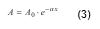
Attenuation

When an ultrasonic wave propagates through a medium, ultrasonic attenuation is caused by a loss of energy in the ultrasonic wave and other reasons. The attenuation can be seen as a reduction of amplitude of the wave. There are some factors affecting the amplitude and waveform of the ultrasonic wave, such as ultrasonic beam spreading, energy absorption, dispersion, nonlinearity, transmission at interfaces, scattering by inclusions and defects, Doppler effect and so on. To characterize the ultrasonic attenuation quantitatively, attenuation coefficient α is defined as follows:


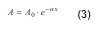
where A is the peak amplitude of the wave at propagation distance x, A0 is the initial peak amplitude.The attenuation coefficient α is experimentally determined from the variation of the peak amplitude with the propagation distance, and it can be given in decibel per metre (dB/m) or in neper per metre (Np/m). In general, the attenuation coefficient highly depends on frequency. Since this frequency dependence reflects microstructures of materials, it can be used for characterizing microscopic material properties relating to chemical reactions and mechanical processes.

Wavelength


Wavelength λ is the distance over which one spatial cycle of the wave completes and the following expression can be given,


where v is the ultrasonic velocity and f is the frequency. Wavelength is a useful parameter in ultrasonic sensing and evaluations. In ultrasonic detection of a small object, the smallest size that can clearly be detected must be larger than half a wavelength at the operating frequency. If the critical size of an object to be detected is known, such prior information on size is helpful for selecting an appropriate frequency for measurements.

Reflection and Transmission


When an ultrasonic wave perpendicularly impinges on an interface between two
media as shown in Fig. 2, a part of the wave is reflected back to the medium 1 and the remainder is transmitted to the medium 2.


The ratio of the amplitude of the reflected wave AR to that of the incident wave AI is called reflection coefficient R, and the ratio of the amplitude of the transmitted wave AT to that of the AI is called transmission coefficient T. Considering a balance of stresses and a continuity of velocities on both sides of the interface, the reflection and transmission coefficients, R and T can be given as follows:


where subscripts 1 and 2 refer to the medium 1 and 2, respectively, and z is the acoustic impedance defined as (1). It can be seen from these equations that the maximum transmission of ultrasonic wave occurs when the impedances of the two media are identical, and most of ultrasonic wave is reflected when the two media have very different impedances. The reflection and transmission at interface play an important role in designing ultrasonic sensing systems and understanding experimental results with the ultrasonic systems.

Refraction and Mode Conversion

When an ultrasonic wave obliquely impinges on an interface between two media as shown in Fig. 3, several things happen depending on the incident angle of the wave as well as the material properties of the two media. One of important things is refraction in which a transmitted wave has a different angle from the incident. The refraction is basically caused by the velocity difference on either side of the interface. The refraction angle can be calculated from Snell’s law if the velocities of the two media and the incidence angle are known.


Another important phenomenon is mode conversion that is a generation of one type of wave from another type in refraction as shown in Fig. 3. For example, a longitudinal wave incident on an interface between liquid and solid is transmitted partially as a refracted longitudinal wave and partially as a mode converted shear wave in the solid. Mode conversion can also take place on reflection if the liquid shown in Fig. 3 is a solid. It is noted that any types of waves can be converted to another type, e.g. from a shear wave to a longitudinal wave, and from a longitudinal wave to a surface wave. The angles of reflection and/or refraction by mode conversion can be calculated fromSnell’s law.

Figure 4 shows a simulation result for refraction and mode conversion, calculated by a finite difference method. We can see that an incident plane wave (longitudinal wave) of 10 in water is refracted at the refraction angle of 43 in steel and simultaneously converted to shear wave at refraction angle of 22.


Ultrasonic Reference:


Wave Motion in Elastic Solids
Wave Motion in Elastic Solids 

Piezoelectric and Acoustic Materials for Transducer Applications
Piezoelectric and Acoustic Mater...
 

Can't find what you looking for? Please search in the web here:


Custom Search

Applications of Ultrasound

Piezoelectric Biosensor

Friday, November 19, 2010


Biosensors are chemical sensors that take advantage of the high selectivity and sensitivity of a biologically active material. It is well known that the resonant frequency of an oscillating piezoelectric crystal can be affected by a change in mass at the crystal surface. Piezoelectric immunosensors are able to measure a small change in mass.

A biosensor is an analytical tool consisting of biologically active material used in close conjunction with a device that will convert a biochemical signal into a quantifiable electrical signal. Biosensors have many advantages, such as simple and low-cost instrumentation, fast response times, minimum sample pretreatment, and high sample throughput. Although biosensors are beginning to move toward field testing and commercialization in the United States, Europe, and Japan, relatively few have been commercialized. Increased research in this area demands the development of novel materials, new and better analytical techniques, and new and improved biosensors. Some potential applications of biosensors are agricultural, horticultural and veterinary analysis; pollution, water and microbial contamination analysis; clinical diagnosis and biomedical applications; fermentation analysis and control; industrial gases and liquids; mining and toxic gases; explosives and military arena; and flavors, essences and pheromones.
 
A biosensor has two components: a receptor and a detector. The receptor is responsible for the selectivity of the sensor. Examples include enzymes, antibodies, and lipid layers. The detector, which plays the role of the transducer, translates the physical or chemical change by recognizing the analyte and relaying it through an electrical signal. The detector is not selective. For example, it can be a pH-electrode, an oxygen electrode or a piezoelectric crystal. Figure 1 describes a typical biosensor configuration that allows measurement of the target analyte without using reagents. 

The device incorporates a biological-sensing element with a traditional transducer. The biological-sensing element selectively recognizes a particular biological molecule through a reaction, specific adsorption, or other physical or chemical process, and the transducer converts the result of this recognition into a usable signal, which can be quantified. Common transduction systems are optical, electro-optical, or electrochemical; this variety offers many opportunities to tailor biosensors for specific applications. For example, the glucose concentration in a blood sample can be measured directly by a biosensor (which is made specifically for glucose measurement) by simply dipping the sensor into the sample.

Two classes of bio-recognition processes-bio-affinity recognition-and bio-metabolic recognition, offer different methods of detection. Both processes involve the binding of a chemical species with another, which has a complementary structure. This is referred to as shape-specific binding. In bio-affinity recognition, the binding is very strong, and the transducer detects the presence of the bound receptor-analyte pair. The most common types of processes are receptor-ligand and antibody-antigen binding. 

In bio-metabolic recognition, the analyte and other co-reactants are chemically altered to form the product molecules. The biomaterials that can be recognized by the bio-recognition elements are as varied as the different reactions that occur in biological systems. Table 1 lists a number of common analytes that could prove attractive for developing biosensors of appropriate specificity and sensitivity. 


Almost all types of biological reactions, (chemical or affinity), can be exploited for biosensors. The concept of shape-specific recognition is commonly used to explain the high sensitivity and selectivity of biological molecules, especially antigen-antibody systems. The analyte molecule has a complementary structure to the antibody, and the bound pair is in a lower energy state than the two separate molecules. This binding is very difficult to break. Table 2 summarizes a variety of biosystem-transducer combinations in terms of transducer, measurement mode and potential application.
 

The interaction of antibodies with their corresponding antigens is an attractive reason for attempting to develop antibody-based chemical biosensors, i.e. immunosensors. Theoretically, if an antibody can be raised against a particular analyte, an immunosensor could be developed to recognize it. Despite the high specificity and affinity of antibodies towards complementary ligand molecules, most antibody-antigen interactions do not cause an electronically measurable change. However, the remarkable selectivity of antibodies has fueled much research to overcome this intrinsic problem. The piezoelectric effect in various crystalline substances is a useful property that leads to the detection of analytes. Figure 2 shows a schematic diagram of an immunosensor device.

 The piezoelectric immunosensor is thought to be one of the most sensitive analytical instruments developed to date, being capable of detecting antigens in the picogram range. Moreover, this type of device is believed to have the potential to detect antigens in the gas phase as well as in the liquid phase.

A piezoelectric sensor that could reliably detect the mycobacterial antigen in biological fluids would be of enormous use. For instance, detection of the antigen in saliva could constitute a noninvasive method of screening high-risk populations. One tested piezoelectric crystal sensor gives results within a couple of hours after exposing the electrode to a liquid containing the antigen. The apparatus would be quite portable, so the immunological tests could be performed virtually anywhere, and the results could be obtained very quickly. 

Theoretical Principals


The basic equations describing the relationship between the resonant frequency of an oscillating piezoelectric crystal and the mass deposited on the crystal surface have been derived by Sauerbrey, Stockridge, and Lostis. Each followed a different path, but their final equations are similar, the Sauerbrey equation being the most widely accepted. In 1959, Sauerbrey developed an empirical equation for AT-cut quartz crystals vibrating in the thickness shear mode that describes the relationship between the mass of thin metal films deposited on quartz crystals and the corresponding change in resonant frequency of the crystal:
(1)

where, DF = frequency change in oscillating crystal in Hz, F = frequency of piezoelectric quartz crystal in MHz, DM = mass of deposited film in g, and A = area of electrode surface in cm2.

These relationships not only apply to film deposition but also to particulate deposition. When vibrating in the thickness-shear mode, the oscillating frequency of an AT-cut quartz crystal is given by: 

(2)
where F is the frequency of the crystal, N is the material constant (N = 1.66 MHz-mm for AT-cut quartz crystal), and a is the thickness of the crystal plate. Thus,


(3)
 for finite amount of change, we may write
(4)
Dividing Equation 4 by Equation 2 gives:
(5)
If the thickness is defined as
(6)
where M is the mass of the electrically driven portion of the crystal, A is the area of the electrically driven portion of the crystal, and r is the density of the crystal.

Assuming constant density, we have:
(7)
If the changes are finite,
(8)
Using Equation 6 yields
(9)
Thus,
(10)
Since F and M are constants, we may say:
DF = -k DM
(11)
The oscillating frequency of the crystal changes linearly with the change in mass on the crystal. The mass change occurs due to deposition of materials on the surface of the crystal. This relationship is only valid for small mass changes. For larger mass changes, it would be invalid since the density would change.

Equation 10 may also be stated as:
(12)
This shows that the term
 D
D
M
    
would increase if the base oscillating frequency of the crystal is increased. The term itself is the sensitivity of the crystal sensor, and Equation 12 shows that sensitivity is directly proportional to the base frequency of the crystal.

If a gas stream is sent flowing over the surface of the piezoelectric crystal and if it contains an analyte with concentration C, then
(13)
where C is the concentration of the analyte in the gas stream, DM is the mass of analyte in the gas stream, and V is the volume of the gas in the stream. The volume of the gas stream is related to the sampling time by the following relationship:

V = q · t
(14)
where q is the flow rate of the gas stream, and t is the sampling time. Equation 13 can be rewritten as:
(15)
where E is the collection efficiency of the coating material. If E is assumed to be equal to one and substituting Equation 15 into Equation 12, we may say
(16)
or
(17)
Thus, the change in the oscillating frequency of the crystal is related to both the sampling time and the concentration of the particles in the carrier gas. If we keep the sampling time, career gas flow rate and the mass of the analyte in the gas stream constant, we may restate Equation 17 as
DF = K C
(18)
Equation 18 shows that the change in frequency of the crystal is directly proportional to the concentration of the analyte in the gas stream flowing over it.

The piezoelectric crystal detector can be a very powerful analytical tool because of the relationship shown for the change in frequency to the analyte concentration with high sensitivity. Conversely, the above explanation shows that the crystal detector indiscriminately changes frequency due to the deposition of mass of any material on its surface. Thus, it is the task of the researcher to choose a coating that will undergo a highly selective chemical or physical binding with the substance to be detected. Only then can a highly selective sensor be constructed that will be sensitive to the subject to be detected.


Surface Plasmon Resonance Based Sensors (Springer Series on Chemical Sensors and Biosensors)Handbook of Biosensors and Biosensor KineticsHydrogel Sensors and Actuators: Engineering and Technology (Springer Series on Chemical Sensors and Biosensors)
Surface Plasmon Resonance Based Sensors (Sp...
$179.00
Handbook of Biosensors and Biosensor Kinetics
by Ajit Sadana
$590.00
Hydrogel Sensors and Actuators: Engineering...
$66.30
Nanomaterials for Biosensors (Nanotechnologies for the Life Sciences)Smart Biosensor Technology (Optical Science and Engineering)Nanostructured Materials for Electrochemical Biosensors (Nanotechnology Science and Technology Series)
Nanomaterials for Biosensors (Nanotechnolog...
$174.54
Smart Biosensor Technology (Optical Science...
$162.95
Nanostructured Materials for Electrochemica...
$89.00